Altered expression of Nurr1 in mice
lacking dopamine D2 receptor
Thesis by
Kyou Chan Choi
Department of Medical Science
Altered expression of Nurr1 in mice
lacking dopamine D2 receptor
Directed by Professor Jong-Eun Lee
The Master's Thesis
submitted to the Department of Medicine Science
the Graduate School of Yonsei University
in partial fulfillment of the requirements for the
degree of Master of Medical Science
Kyou Chan Choi
This certifies that the Master's Thesis
of Kyou Chan Choi is approved.
---
[Thesis Supervisor : Jong-Eun Lee]
---
[Thesis Committee Member]
---
[Thesis Committee Member]
The Graduate School
Yonsei University
Acknowledgments
지도와 지도와 지도와 지도와 관심을관심을 보여주셨던관심을관심을 보여주셨던보여주셨던 지도보여주셨던 지도지도지도 교수님인교수님인 이종은교수님인교수님인 이종은이종은이종은 교수님께교수님께교수님께 먼저교수님께 먼저먼저먼저 감사를 감사를 감사를 감사를 전합니다전합니다전합니다전합니다. . . . 아울러아울러아울러아울러 2 2 2 2년여년여 동안년여년여 동안동안동안 항상항상항상항상 많은많은 지도와많은많은 지도와지도와지도와 관심으로관심으로관심으로관심으로 저를 저를 저를 저를 일깨워주셨던일깨워주셨던일깨워주셨던 백자현일깨워주셨던 백자현백자현백자현 교수님과교수님과 논문교수님과교수님과 논문논문 심사위원으로논문 심사위원으로심사위원으로심사위원으로 수고해주신수고해주신수고해주신수고해주신 김찬형 김찬형 김찬형 김찬형 교수님께도교수님께도교수님께도교수님께도 감사의감사의감사의감사의 말씀말씀말씀말씀 드립니다드립니다드립니다드립니다. . 항상. . 항상항상항상 함께함께함께 하면서함께 하면서하면서하면서 가족처가족처가족처가족처 럼 럼 럼 럼 대해주셨던대해주셨던 실험실대해주셨던대해주셨던 실험실실험실실험실 동료인동료인동료인동료인 신승우신승우, 신승우신승우, , , 김김김규석김규석, 규석규석, , , 박은아박은아박은아, 박은아, 강은영, , 강은영강은영강은영, , , , 리명리명리명리명 철 철 철 철, , 김성렬, , 김성렬김성렬김성렬, , 이상협, , 이상협이상협 선생님께이상협 선생님께선생님께 감사를선생님께 감사를 전합니다감사를감사를 전합니다전합니다전합니다. . . . 그리고그리고그리고그리고 지금은지금은지금은지금은 다른다른다른다른 곳에서 곳에서 곳에서 곳에서 각자의각자의각자의 일에각자의 일에일에일에 매진하고매진하고매진하고 있을매진하고 있을있을 안연희있을 안연희안연희안연희, , 이일선, , 이일선이일선, 이일선, , , 김명환김명환, 김명환김명환, , , 배미현배미현배미현, 배미현, , , 김용년 김용년 김용년 김용년, , , 김성재, 김성재김성재, 김성재, 김량여, , 김량여김량여김량여, , , 윤성환, 윤성환, 윤성환윤성환, , , 김청섭김청섭김청섭 선생님께도김청섭 선생님께도선생님께도선생님께도 감사를감사를감사를감사를 드립니드립니드립니드립니 다 다 다 다. . . 아울러. 아울러아울러 언제나아울러 언제나언제나언제나 많은많은많은 격려를많은 격려를 해주셨던격려를격려를 해주셨던해주셨던해주셨던 김종렬김종렬김종렬김종렬, , 김한조, , 김한조김한조 선생님께김한조 선생님께선생님께선생님께 감사를 감사를 감사를 감사를 드립니다드립니다드립니다. 드립니다. . . 학창시절의 학창시절의 학창시절의 학창시절의 즐거움을즐거움을 함께즐거움을즐거움을 함께함께 한함께 한한한 태석태석, 태석태석, , 석현, 석현석현석현 선배와선배와 형석선배와선배와 형석형석형석, , , , 석호석호, 석호석호, , 호중, 호중호중호중, , , , 준영 준영 준영 준영, , , 웅희, 웅희웅희웅희, , , 상우, 상우에게상우상우에게에게 감사를에게 감사를감사를감사를 전합니다전합니다. 전합니다전합니다. . . 또한또한또한또한 십년이십년이 넘게십년이십년이 넘게넘게넘게 한결한결한결 같은한결 같은같은같은 친구로 친구로 친구로 친구로 남아준남아준남아준 혜원남아준 혜원혜원혜원, , , , 지호지호지호지호에게에게 진심으로에게에게 진심으로진심으로진심으로 고마움을고마움을고마움을고마움을 전합니다전합니다. 전합니다전합니다. . 언제나. 언제나언제나 언제나 사랑이란 사랑이란 사랑이란 사랑이란 이름으로이름으로이름으로 저를이름으로 저를저를 감싸주셨던저를 감싸주셨던감싸주셨던 부모님감싸주셨던 부모님부모님, 부모님, 누나, , 누나누나누나,,,, 매형매형매형, 매형, , 사랑하는, 사랑하는사랑하는사랑하는 나의 나의 나의 나의 가족들에게가족들에게가족들에게가족들에게 감사를감사를감사를감사를 전하면서전하면서 이전하면서전하면서 이이이 논문을논문을논문을 마칩니다논문을 마칩니다마칩니다마칩니다.... 200 200 200 200444년4년년년 12 12 12 12월월월 월 최 최 최 최 규규규규 찬찬찬 찬ii
Contents
Abstract……….……...………….……….1 Ⅰ Ⅰ Ⅰ Ⅰ. Introduction……...….………..……..………….………3 Ⅱ Ⅱ Ⅱ Ⅱ. Materials and Methods……….………..71. Animals and dissection.………..………7
2. Mesencephalic and striatal culture ………….……….7
3. Immunocytochemistry……….………..9
4. RT-PCR reaction………10
5. Cell Culture………...11
6. Transfection and Luciferase reporter gene assay…………..11
Ⅲ Ⅲ Ⅲ Ⅲ. Results………12
1. The Role of D2R in cell death and development …..……….12
2. Nurr1 mRNA expression during midbrin development in WT and D2R-/- mice……….19
3. Transactivation of Nurr1 by dopamine D2 receptor....….…22
Ⅳ Ⅳ Ⅳ Ⅳ. Discussion………..……….26 Ⅴ Ⅴ Ⅴ Ⅴ. Conclusion………..…29 References………..……..……31
iii
List of Figures and Table
Figure 1. The representative Photomicrographs of TH-positive neurons in ventral mesencephalic-striatal co-culture on day 8………...…………..……….16
Figure 2. TH staining in mouse mesencephalic culture.……….….15
Figure 3. TH staining in mouse mesencephalic-striatal co-culture16
Figure 4. Nurr1 mRNA expression during midbrin development in WT and D2R-/- mice………...20
Figure 5. Immunohistochemistry for Nurr1 on coronal sections of ventral midbrain in WT and D2R-/- mice...21
Figure 6. Transactivation of Nurr1 by dopamine D2 receptor...24
Table1. Estimated EC50 values of dopamine for D2 receptor in HEK293 cells………....25
1
ABSTRACT
Altered expression of Nurr1 in mice
lacking dopamine D2 receptor
Kyou Chan Choi
Department of Medical Science
The Graduate School, Yonsei University
(Directed by Professor Jong-Eun Lee)
The dopamine D2 receptor (D2R) is highly expressed in the central nervous system and is crucial for the regulation of various neurophysiological functions. In mice lacking D2R (D2R -/-), the number of mesencephalic tyrosine hydroxylase (TH)-positive at embryonic stage was significantly low, when compared to that of wild-type (WT) mice, indicating an alternation in dopaminergic (DAergic) neuronal development in the absence of D2R. I have
2
investigated the expression of Nurr1, an orphan nuclear receptor is known to be essential for development of midbrain DAergic cells. RT-PCR and immunohistochemical anlaysis revealed that Nurr1 expression was selectively decreased in D2R -/- mice at embryonic stage, which was rescued in the adulthood. These data suggest that D2 receptor-mediated signaling is important in the development of midbrain do DAnergic neurons and probably in association with Nurr1.
Key words: Dopamine D2 receptor, Nurr1, Dopaminergic neuron, Differentiation
3
Altered expression of Nurr1 in mice
lacking dopamine D2 receptor
(Directed by Professor Jong-Eun Lee)
Department of Medical Science
The Graduate School, Yonsei University
Kyou Chan Choi
Ⅰ
Ⅰ
Ⅰ
Ⅰ. Introduction
Dopamine (DA) is the predominant catecholamine neuro-transmitter in the mammalian brain, where it controls diverse functions such as locomotor activity, neuroendocrine hormone release1. DA is synthesized from tyrosine hydroxylase (TH), the rate limiting enzyme in the catecholamine biosynthesis. The
4
majority of neurons that produce dopamine originate in the ventral midbrain in the substantia nigra (A9) and the ventral tegmental area (A10). The importance of proper DAergic system function is
evident when any of these systems become compromised such as in Parkinson’s disease2, Schizophrenia3. The mechanism of DAergic neuronal development might be useful in directing commitment of DAergic neuronal cells in vitro before grafting in therapies for Parkinson’s disease21,22,23. It is known that factors are often critical as components of developmental regulator, and transcription factors such as Nurr18,9, Ptx329,30,31, and Lmx1b32 were known to be critical in development of DAergic neuron .
The transcription factor Nurr1 is an orphan member of the nuclear steroid/thyroid hormone receptor superfamily, which is expressed predominantly in the central nervous system4.
Genetically modified mice lacking both copies of the Nurr1 gene fail to generate mesencephalic neurons7,8. The exogenous expression of Nurr1 into stem cells enhances differentiation and maturation into DAergic neurons. Recent in vitro studies showed that Nurr1 is able to activate TH gene transcription9. Despite these interesting findings, little is known about the mechanisms that
5
regulate Nurr1 expression in DAergic neurons.
Nurr1 can also activate gene transcription independent of ligands, possibly being influenced by other signaling pathways acting via cell-membrane-bound receptors.
DA acts through membrane receptors of the seven
transmembrane domain G-protein coupled receptor family. Molecular cloning of the dopamine receptor family revealed five receptor subtypes, D1 through D5. They have been classically divided into two groups based on ligand specificity and effector coupling. The D1-like receptors, comprising D1 and D5 receptors, are positively coupled to adenylyl cyclase by the G protein Gs, whereas the D2-like receptors, comprising D2, D3 and D4 receptors, whose activation results in inhibition of adenylyl cyclase and suppression of cAMP production11,12. The D2 subtype of DA receptor represents the main autoreceptor of the DAergic system16,17,18.
Recently, it has been reported that the signaling through D2 receptor (D2R) was found to be crucial for anti-proliferative effects and cell death in pituitary tumor cells13. Indeed, it was found that the DAergic agonists could induce the anti-proliferative effects and
6
cell death via D2R. I investigated that how the absence of D2R affects the neuronal cell death and development in mice lacking D2R (D2R -/-). I found that in the absence of D2R, development of DAergic neuron was blunted in association with altered expression of Nurr1 in these mice.
7
Ⅱ
Ⅱ
Ⅱ
Ⅱ. Materials and Methods
1. Animals and dissections
The D2R-/- mice and wild type littermates originated from the mating of heterozygous D2R-/- mice identified by Southern hybridization analyses as described by Baik et al16. The D2R-/- mice had a mixed 129SV/C57BL/6 genetic background, with a 75% constribution of C57BL/6. Heterozygous D2R-/- mice were mated from 18:00h to 8:00h. Insemination was confirmed by vaginal plug and considered as E0. Pregnant mothers were killed in accordance with Society for Neuroscience guidelines. At E15 days, embryos were excised, brains were removed from the skull and the striatum and the ventral mesencephalon were dissected by mild mechanical trituration in ice-cold Ca++ and Mg++ free Hank`s balanced salt solution with a supplement of 4.2 mM sodium bicarbonate.
2. Mesencephalic and striatal culture
Cells were dissociated by mechanically dispersing tissue pieces with a 1 ml pipet. Subsequently, cells were centrifuged at 200 g for 5min and resuspended in Minimum essential medium (MEM, SIGMA) containing 2 mM L-glutamine, 1 g/L glucose, 5 % heat
8
inactivated fetal bovine serum, 5 % heat inactivated horse serum. Dissociated cells were plated in 35 mm culture dishes onto pre-coated 24 mm glass cover slips with 0.1 mg/ml ploy-D-lysine (SIGMA) and 2 µg/ml laminin (SIGMA). After 2 days in cultures, 5 µM cytosine β-D-arabinofuranoside (SIGMA) was added to inhibit glial cell proliferation. Cultures were maintained at 37 ℃℃℃℃ in a humidified atmosphere of 5 % CO2 and 95 % air for 6 days. The Exposure to MPP+ was begun on Day 7 in vitro. The medium was removed and replaced with serum-free growth medium. MPP+I-(Research Biochemicals, Inc.) was dissolved in culture medium and added at the concentration specified for 24 hr.
3. Immunocytochemistry
The cultures were processed for TH immunocytochemistry using the biotin-avidin peroxidase method. Cultures grown on cover slips were rinsed with 0.1 M phosphate buffered saline (PBS, pH 7.4) and fixed for 30 min with 4 % paraformaldehyde in 0.1 M PBS at 4 ℃℃℃℃ overnight. After washing (3X) with PBS, the fixed cultures were treated for 30 min with blocking solution (1 % BSA, 0.2 % Triton X-100, and 10 % normal serum in PBS). The cultures were
9
then incubated overnight at 4 ℃℃℃℃ with a monoclonal antibody against TH (1:1000, Diasorin Stillwater, MN, USA). After washing, the cells were exposed to a biotinylated anti-mouse IgG (1:200, Vector lab.) for 1 hr at room temperature. Cultures were rinsed with PBS, then treated for 1 hr at room temperature with avidin-biotinylated horseradish peroxidase complex (Vectastain Elite ABC kit, Vector lab. Burlingame, CA) The peroxidase was visualized with diaminobenzidine (DAB) and hydrogen peroxide. Specimens were dehydrated through a graded ethanol series and mounted on slide glasses with Permount®®®® solution (Fisher Scientific, New Jersey, USA). The number of TH-positive neurons were evaluated under a microscope and counted in 20 randomly selected fields at 100 X magnification. A 2.5 mm2 square grid was inserted into the microscopic field.
For immunohistochemical analysis of tissues, brains were removed, rinsed in ice-cold PBS fixed 4% paraformaldehyde, cryoprotected in 30% sucrose in 0.1 M PBS (pH 7.4) overnight at 4℃℃℃℃,,,, placed in OCT Tissue Tek embedding medium (Sakura®, CA, USA) and frozen on dry ice. Thin section (10㎛㎛㎛) were cut on a ㎛
10
immunohistochemistry.
4. RT-PCR reaction Tissues were removed from freshly killed mice and homogenized
by polytron homogenizer and lysis buffer containing 3 M LiCl2, 6M urea, 0.1 % SDS, 10 mM NaOAc (pH 5.0), and 0.2 mg/ml heparin. After overnight precipitation at 4℃℃℃℃, the precipitates was washed with buffer containing 4 M LiCl2 , 8 M urea and 10 mM NaOAc (pH 5.0) and then extracted with acidic phenol. Total RNA was prepared from isolated mesencephalon of mice brain using LiCl RNA extraction buffer. First strand cDNAs were generated from total RNA using reverse-transcription with random primer by denaturing at 90 ℃℃℃℃ for 4 min, annealing at room temperature for 10 min and extending at 42 ℃℃℃℃ for 50 min. The following primers
were used to amplify target cDNA: Nurr1,
5´TAAAAGGCCGGAGAGGTCGTT3´,5´CTCTCTTGGGTTCCT
TGAGCC3´ β-actin, 5´GATGACGATATCGCTGCGCT3´,
5´GCTCATTGCCGATAGTGATGACCT3´. Conditions for PCR amplifications were as follows: 94 ℃℃℃℃ for 5 min, 30 cycles at 94 ℃℃℃℃ for 1 min, 60 ℃℃℃℃ for 1 min, 72 ℃℃℃℃ for 1 min, and final extension at
11
72 ℃℃℃℃ for 7 min. The PCR reaction products were run on 1.5 % agarose gels containing ethidium bromide (0.5 µg/ml), to mark and visualize the PCR products.
5. Cell Culture
HEK (Human Embryonic Kidney) 293 cells were grown in
Dulbecco’s modified Eagle’s medium (DMEM) (Gibco,
Gaithersbrug, Maryland, USA) supplemented with 10% fetal bovine serum, penicillin G (100 units/ml), streptomycin sulfate (100
µg/ml) and amphotericin B (250mg/ml).
6. Transfection and Luciferase reporter gene assay
HEK (Human Embryonic Kidney) 293 cells were grown to confluence in 6-well plates. Transient transfections of the HEK293
were performed using PEI (polyethylenimine)-mediated
transfection reagent, jetPEI™ (Qbiogene, Inc., Carlsbad, CA, USA). Briefly, 70% ~ 80% confluent monolayers in 6-well plates were incubated at 37°C with transfection mixture containing 1.5 µg of D2R, 1.5 µg of Nurr1, 1.5 µg of a reporter plasmid regulated by three copies of the NurRE (Nur response element), 0.5 µg of
12
pCH110 carrying the β-galactosidase gene. After 3 h, the transfection mixture was replaced with fresh growth medium. Assays were performed 48 h after transfection. Cells were preincubated overnignt in serum free DMEM medium before cells were treated with dopamine. The cells were pretreated with Haloperidol (1µM) for 5min or Pertussis toxin (100ng/ml) for 12h, followed by treatment with various concentrations of dopamine for a further 6 hr at 37 °C. HEK293 cells were assayed for luciferase activity using the luciferase assay system (Promega), and luminescence was measured using a 96-well luminometer (Microlumat; EG & Berthold, Bad Wilbad, Germany). The expression of the reporter gene was normalized using β-galactosidase activity19,20. Transfection in the control group was performed under the transfection conditions above with 1.5 µg of D2R, 1.5 µg of Nurr1, 1.5 µg of a reporter plasmid regulated by three copies of the NurRE (Nur response element), 0.5 µg of pCH110 carrying the β-galactosidase gene, but without stimulating by dopamine. The mean values of the data obtained were fitted to a sigmoid curve with a variable slope factor using the nonlinear squares regression in a GRAPHPAD PRISM®. EC50 (nM) values
13
were described as mean± S.E. All of the luciferase reporter gene activity assays were performed at least four independent.
14
Ⅲ
Ⅲ
Ⅲ
Ⅲ. Results
1. The Role of D2R in cell death and development
To investigate the regulation of DAergic neuronal cell death and development in WT and D2R-/- mice, I used primary neuronal cells of the mesencephalon from WT and D2R-/- mice. The selective neurotoxin 1-methyl-1,2,3,6-tetra-hydropyridine (MPTP) has been widely used to generate animal models of Parkinson’s disease. When administered in vivo, MPTP crosses the blood–brain barrier and is converted, mainly in glial cells, into its effective form, 1-methyl-4-phenylpyridinium (MPP+), by monoamine oxidase B33. MPP+ finally enters mitochondria by an energy-dependent mechanism inhibiting the activity of this organelle and leading to a drop in cellular ATP levels and subsequent DAergic neuronal cell death34.
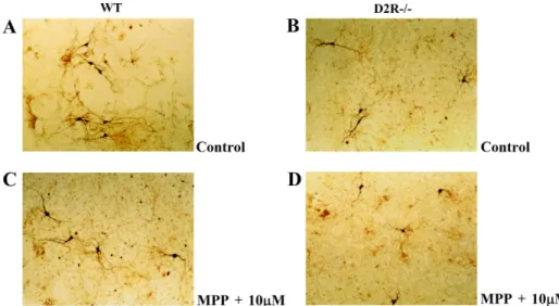
TH immunohistochemistry showed that treatments of MPP+ (10
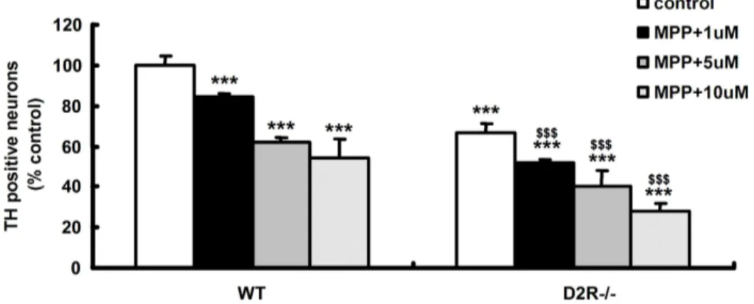
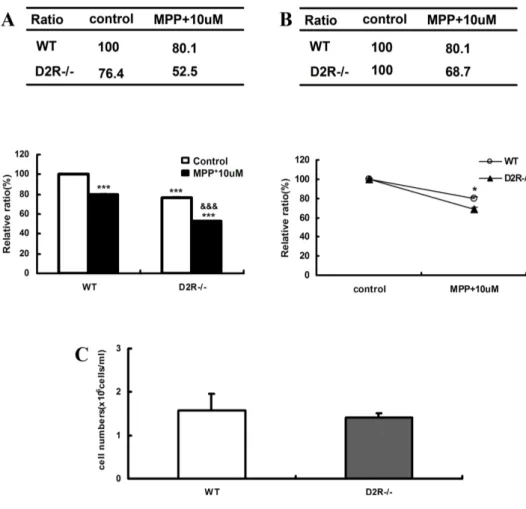
µM) for 24hr induced DAergic neuronal cell death(Fig. 1 C and D). After 24hr exposure of the MPP+ (1-10 µM) to the cells, significant decrease of the DAergic neuronal cells was observed in a dose dependant manner (Fig 2). D2R-/- mice was more susceptible to neurotoxin, when compared to WT (Fig 3B). Interestingly, the
15
number of mesencephalic TH-positive neurons at embryonic stage in D2R-/- mice was significantly low, when compared to that of WT mice (Fig. 2, and 3A). However, comparison of total number of neurons in E15 days mesencephaon of WT and D2R-/- showed no significant differences between the two genotypes (Fig. 3C). Tyrosine hydroxylase (TH) expression level was monitored in SNc (substantia nigra compacta) and VTA of D2R-/- and wild-type (WT) mice. These results suggest that absence of D2Rs may be involved in the development of mesencephalic neuron.
16
Fig 1. The representative Photomicrographs of TH-positive neurons in ventral mesencephalic-striatal co-culture on day 8. (A) Wild-type (WT), control (B) D2R-/- , control (C) Wild-type (WT), exposure to 10 µM of MPP+ for 24hr (D) D2R-/-, exposure to 10 µM of MPP+ for 24hr.
17
Fig 2. TH staining in mouse mesencephalic culture. TH+ cells counting in mesencephalic cultures established from WT and D2R-/- E15 days mice after treatment with MPP+ (1-methyl-4-phenylpyridinium). WT (n=5) and D2R-/- (n=7) embryonic mice that were treated 1-10 µM with MPP+ (24hr exposure) were performed by immunocytochemistry. * p < 0.001 compared wild type control. $ p < 0.001 compared with D2R-/- control.
18
Fig 3. TH staining in mouse mesencephalic-striatal co-culture. (A), (B) TH-positive cells counting in mesencephalic-striatal co-cultures established from wild-type (WT) and D2-/- E15 days mice after treatment with MPP+ (1-methyl-4-phenylpyridinium). WT (n=4) and D2R-/- (n=5) embryonic mice that were treated with 10 µM MPP+ (24hr exposure) were performed by immunocytochemistry. * p < 0.001 compared wild type control. $ p < 0.001 compared with D2R-/- control. (C) The number of mesencephalic neurons of E15 days WT and D2R-/- mice.
19
2. Nurr1 mRNA expression during midbrin development in WT and D2R-/- mice.
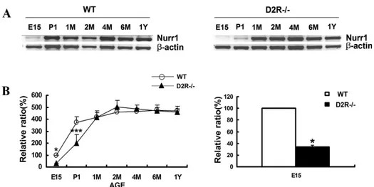
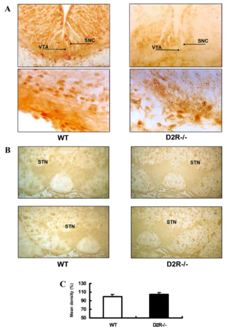
As mentioned above, the transcription factor/nuclear receptor Nurr1 is essential for the development of mesencephalic DAergic neuron7,8. Nurr1 mRNA is expressed in substantia nigra pars compacta (SNC) and the ventral tegmental area (VTA). By using semiquantitative RT-PCR and immunohistochemistry, I analysed the Nurr1 gene expression during ontogeny of the mesencephalon. RT-PCR showed that Nurr1 expression was selectively decreased in D2R -/- mice at embryonic stage and was rescued in the adulthood (Fig 4). From embryonic 15 days to post-natal 1day, Nurr1 immunoreactivity was selectively decreased in D2R-/- mice. However in the adult, there was no obvious difference in the Nurr1 immunoreactivity of ventral tegmental area (VTA) in WT and D2R-/- mice (Fig 5B and C). These datasuggest that in the absence of D2R, the development of DAergic neuron is blunted in association with altered expression of Nurr1.
20
Fig 4. (A) Developmental expression of Nurr1 mRNA in wild-type (WT) and D2R-/- mice by RT-PCR analysis. Total RNAs from midbrain of WT and D2R-/- mice were analyzed by reverse transcription PCR (RT-PCR) for Nurr1 transcripts. RT-PCR analysis was performed at various stage of development. The developmental stages analyzed were embryonic 15 days (E15), postnatal 1 day (P1), M (1, 2, 4, 6 months) and Y (1 year). * p < 0.001 compared with wild-type E15 days. (B) Data were plotted (in %) for Nurr1 mRNA levels, respectively, in relation to mRNA levels of the β-actin gene, as an internal standard.
21
Fig 5. Immunohistochemistry for Nurr1 on coronal sections of ventral midbrain in WT and D2R-/- mice. (A) Nurr1 immunoreactivity in ventral tegmental area (VTA) of E15 days in WT and D2R-/- mice. (B) Nurr1 immunoreactivity in ventral tegmental area (VTA) of adult in WT and D2R-/- mice. (C) The relative Nurr1 immunoreactivity in ventral tegmental area (VTA) of adult in WT and D2R-/- mice.
22
3. Transactivation of Nurr1 by dopamine D2 receptor.
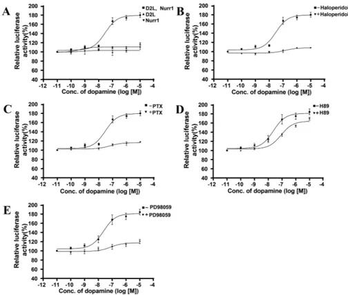
To investigate whether the activation of Nurr1 is involved in signaling pathway mediated by D2R, the DNAs encoding D2R, Nurr1, and a reporter plasmid regulated by three copies of the NurRE (Nur response element) were transiently transfected into HEK 293 cells. When increasing concentrations dopamine were treated, a typical dose-dependant and saturable induction of luciferase activity was observed. However, I detected no significant effect of dopamine on cells transfected with either D2R or Nurr1 alone (Fig 6A). To confirm activation of Nurr1 is specific to the stimulation of D2R, the cells were pretreated with D2R antagonist, haloperidol for 5 min, followed by the stimulation of D2R with various concentrations of dopamine. D2R-mediated reporter gene activation was completely inhibited by the treatment of haloperidol (Fig 6B), suggesting that the activation of Nurr1 is mediated by stimulation of D2R.
It is now generally accepted that numerous GPCRs can also activate MAPK to influence diverse cellular processes, ranging from the regulation of neuronalsurvival to cell differentiation and
23
predominantly Gß subunit-mediated signaling27. Treatment of PTX caused complete inhibition of Nurr1 activation in HEK 293 cells by stimulation of D2R with DA (Fig 6C), suggestion that the activation of Nurr1 is involved in G protein dependant signaling.
The synthetic compound PD98059 has been characterized as a selective inhibitor of the MAPK pathway by preventing the activation of MEK (MAPK kinase)-128. Pretreatment of cells with 20 μμμμM of PD98059 for 30 min completely inhibit the NurRE-dependent transcriptional activation of luciferase reporter gene by the stimulation of D2R (Fig 6E), suggesting that D2R-medited MAPK activation is essential for the activation of Nurr1.
It was recently suggested that GPCR-mediated activation of induction of Nurr1 in corticotrophs was protein kinsase A (PKA) dependant. The role of PKA on the D2R-mediated activation of Nurr1 was investigated. Treatment of a PKA inhibitor, H-89 did not affect the activation of Nurr1 by the stimulation of D2R (Fig 6D).
These results suggest that the signaling through D2R induces the activation of Nurr1 by PKA-independent and Ras-dependant MAPK pathaway.
24
Fig 6. (A) NurRE-dependent transcriptional activation of luciferase reporter gene upon stimulation of D2R in HEK293 cells. HEK293 cells were transiently transfected with the D2R and Nurr1 together or either Nurr1/ D2R alone followed by the stimulation of D2 receptor with various concentrations of dopamine for 6 h at 37 °C. Effect of dopamine D2 receptor antagonist, Haloperidol (1μM for 5min) (B), (C) an inhibitor of Gi proteins, Pertussis toxin (PTX) (100ng/ml for 12h), (D) PKA inhibitor, H-89 (1μM for 20min), and (E) MAP Kinase Kinase inhibitor, PD98059 (10μM for 30 min) on NurRE-dependent transcriptional activation of luciferase reporter gene upon stimulation of D2R.
25 EC50 Expression/Treatment (nM) D2L, Nurr1 58.8 ± 29.24 D2L only ND Nurr1 only ND +Haloperidol ND +PTX ND +PD98059 ND +H89 75.4 ± 9.67
Table1. Estimated EC50 values of dopamine for D2 receptor in HEK293 cells. EC50 values were determined for the Nur response element luciferase reporter gene activity stimulated by dopamine. Data are mean ±S.E. from at least four independent experiments.
26
. Discussion
Ⅳ
Ⅳ
Ⅳ
Ⅳ
As mentioned above, the mechanism of DAergic neuronal development might be useful in directing commitment of DAergic neuronal cells in vitro before grafting in therapies for Parkinson’s disease21,22,23. It is known that factors are often critical as components of developmental regulator, and transcription factors such as Nurr17,8, Ptx329,30,31, and Lmx1b32 were known to be critical in development of DAergic neuron . The transcription factor Nurr1 is an orphan member of the nuclear steroid/thyroid hormone receptor superfamily, which is expressed predominantly in the central nervous system4. Nurr1 mRNA is expressed across many regions of the developing central nervous system but, in the adult rodent brain, its expression is restricted to the temporal cortex, hippocampus, habenular nuclei, some thalamic nuclei and to DAergic neurons of the substantia nigra pars compacta (SNpc) and ventral tegmental area (VTA)5,6. The onset of Nurr1 expression in the ventral midbrain occurs at embryonic day 10.5 before the appearance of the DAergic marker enzyme, TH, at embryonic day 11.5. Expression of Nurr1 continues in mature DAergic neurons during adulthood, suggesting that Nurr1 may also be required for
27
normal function of mature DAergic neurons.
Little is known about the mechanisms that regulate the development of DAergic neurons and involvement of Nurr1 in this regulation. The importance of Nurr1 in developing and mature DAergic neurons has focused interest on Nurr1 as a potential drug target.
The MAPK signaling cascade is a prominent cellular pathway used by many growth factors, hormones and neurotransmitters to regulate diverse physiological functions25. It has also been observed that the ↓↓↓↓°°°° subunits of G-proteins are able to mediate Ras-dependent MAPK activation26. It has been recently shown that the agonist-stimulated D2R activates MAPK activity Pertussis toxin (PTX) treatment completely revoked stimulation of MAPK mediated by D2R, demonstrating that D2R couple to pertussis toxin-sensitive G proteins in this signaling27.
DA is able to selectively activate some members of the steroid receptor superfamily10.Nurr1 can also activate gene transcription independent of ligands, possibly being influenced by other signaling pathways acting via cell-membrane-bound receptors such as dopamine D2 receptor.
28
These results suggest that signaling through D2R in association with Nurr1 plays a dominant role in development of midbrain DAergic neuron. Future experiments will be required to assess the effect of D2R antagonist, haloperidol, on DAergic neuronal cell death in primary mesencephalic neuron culture system. The identification of interaction between the orphan nuclear receptor Nurr1 and D2R will allow us to elucidate the key molecular mechanism responsible for the DAergic neuronal development and to provide a possible therapeutic strategy to several pathologies with DAergic neuronal degeneration.
29
Ⅴ
Ⅴ
Ⅴ
Ⅴ. Conclusion
This present study showed role of D2R on cell death and development. These results have led me to the following conclusions.
1. TH immunohistochemistry showed that treatments
of MPP+ induced DAergic neuronal cell death. D2R-/- mice was more susceptible to neurotoxin, when compared to WT. The number of TH-positive neurons in ventral tegmental area was significantly low, when compared to that of WT mice.
2. From embryonic 15 days to post-natal 1day, Nurr1 immunoreactivity was selectively decreased in D2R-/- mice. However in the adult, there was no obvious difference in the Nurr1 immunoreactivity of ventral tegmental area (VTA) in WT and D2R-/- mice.
3. Treatment of increasing concentrations dopamine induced a typical dose-dependant and saturable Nurr1 activity, while D2R-mediated reporter gene activation was completely inhibited by the treatment of haloperidol. The activation of Nurr1 is involved in G protein dependant signaling and Ras-MAPK
30
singnaling.
Taken together, these data suggest that absence of D2Rs may be involved in the development of mesencephalic neuron. DA can activate orphan neuclear receptor Nurr1. It may be possible that signaling through D2R in association with Nurr1 plays a dominant role in development of midbrain DAergic neuron.
31
References
(1) Picetti R, Saiardi A, Samad TA, Bozzi, Y, Baik JH, and Borrelli E. Dopamine D2 receptors in signal transduction and behavior. Crit Rev Neurobiol 1997; 11: 121-142.
(2) Lee T, Seeman P, Rajput A, Farley IJ, and hornykiewiez O. Receptor basis for dopaminergic supersensitivity in Parkinson’s disease. Nature 1978; 273: 59-61.
(3) Seeman P. Dopamine receptor and dopamine hypothesis of schizophrenia. Synapse 1987; 1: 133-152.
(4) Law SW, Conneely OM, DeMayo FJ and O’ Malley BW, Identification of a new brain specific transcription factor, Nurr1. Mol. Endocrinol. 1992; 6; 2129–2135.
(5) Saucedo-Cardenas O and Conneely OM, Comparative distribution of Nurr1 and Nurr77 nuclear receptors in the mouse central nervous system. J. Mol. Neurosci. 1996; 7: 51–63.
32
Cellular expression of the immediate early transcription factors Nurr1 and NGFI-B suggests a gene regulatory role in several brain regions including the nigrostriatal dopaminergic system. Mol. Brain Res. 1996; 41: 111–120.
(7) Zetterström RH, Solomin L, Jansson L, Hoffer HJ, Olson L and Perlmann T, Dopamine neuron agenesis in Nurr1-deficient mice. Science 1997; 276: 248–250.
(8) O. Saucedo-Cardenas, Quintana-Hau JD, Le WD, Smidt MP, Cox JJ, F. De Mayo, Burbach JP and Conneely OM, Nurr1 is essential for the induction of the dopaminergic phenotype and the survival of ventral mesencephalic late dopaminergic precursor neurons. Proc. Natl. Acad. Sci. USA 1998; 95: 4013–4018.
(9) K. Sakurada, M. Ohshima-Sakurada, T.D. Palmer and F.H. Gage, Nurr1, an orphan nuclear receptor, is a transcriptional activator of endogenous tyrosine hydroxylase in neural progenitor cells derived from the adult brain. Development 1999; 126: 4017– 4026.
33
(10) Power RF, Lydon JP, Conneely OM and BW O’Malley, Dopamine activation of an orphan of the steroid receptor superfamily. Science 1991; 252: 1543–1546.
(11) Kebabian JW. Dopamine-sensitive adenylyl cyclase: a receptor mechanism for dopamine. Adv Biochem Psychopharmacol 1978; 19: 131-154.
(12) Sibley DR, Monsma FJ Jr. Molecular biology dopamine receptors. Trends Pharmacol Sci 1992; 13: 61-69.
(13) An JJ, Cho SR, Jeong DW, Park KW, Ahn YS and Baik JH. Anti-proliferative effects and cell death mediated by two isoforms of dopamine D2 receptors in pituitary tumor cells. Mol. Cell. Endocrinol. 2003; 206(1-2): 49-62.
(14) Baik JH, Picetti R, Saiardi A, Thiriet G, Dierich A, Depaulis A, LeMeur M and Borrelli E. Parkinsonian-like locomotor impairment in mice lacking dopamine D2 receptors. Nature 1995; 377: 424-428.
34
(15) Wang Z, Benoit G, Liu J, Prasad S, Aarnisalo P, Liu X, Xu H, Walker NP, Perlmann T. Structure and function of Nurr1 identifies a class of ligand-independent nuclear receptors. Nature 2003; 423: 555–560.
(16) L'hirondel M, Cheramy A, Godeheu G, Artaud F, Saiardi A, Borrelli E, Glowinski J. Lack of autoreceptor-mediated inhibitory control of dopamine release in striatal synaptosomes of D2 receptor-deficient mice. Brain Res 1998; 792: 253-262.
(17) Mercuri NB, Saiardi A, Bonci A, Picetti R, Calabresi P, Bernardi G, Borrelli E. Loss of autoreceptor function in dopaminergic neurons from dopamine D2 receptor deficient mice. Neuroscience 1997; 79: 323-327.
(18) Usiello A, Baik JH, Rouge-Pont F, Picetti R, Dierich A, LeMeur M, Piazza PV, Borrelli E. Distinct functions of the two isoforms of dopamine D2 receptors. Nature 2000; 408: 199-203.
35
quantitation of microgram quantities of protein utilizing the principle of protein-dye binding. Anal. Biochem 1976; 72: 248-254
(20) Herbomel P, Bourachot B, Yaniv M. Two distinct enhancers with different cell specificities coexist in the regulatory region of polyoma. Cell 1984; 39: 653-662
(21) Perlow MJ. Brain grafting as a treatment for Parkinson’s disease. Neurosurgery 1987; 20: 335–342.
(22) Date I, Miyoshi Y, Ono T, Imaoka T, Furuta T, Asari S, Ohmoto T, Iwata H. Preliminary report of polymer-encapsulated dopamine-secreting cell grafting into the brain. Cell Transplant 1996; 5 S17–S19.
(23) Kordower JH, Rosenstein JM, Collier TJ, Burke MA, Chen EY, Li JM, Martel L, Levey AE, Mufson EJ, Freeman TB, Olanow CW.
Functional fetal nigral grafts in a patient with Parkinson’s disease: chemoanatomic, ultrastructural, and metabolic studies. J Comp Neurol 1996; 370: 203–230.
36
(24) Kim JH, Auerbach JM, Rodriguez-Gomez JA, Velasco I, Gavin D, Lumelsky N, Lee SH, Nguyen J, Sanchez-Pernaute R, Bankiewicz K, McKay R. Dopamine neurons derived from embryonic stem cells function in an animal model of Parkinson's disease. Nature 2002; 418: 50-56.
(25) Seger R and Krebs EG. The MAPK signaling cascade. FASEB 1995;9: 726-735
(26) van Biesen T, Hawes BE, Luttrell DK, Krueger KM, Touhara K, Porfiri E, Sakaue M, Luttrell LM, Lefkowitz RJ. Receptor-tyrosine-kinase and G beta gamma-mediated MAP kinase activation by a common signalling pathway. Nature 1995; 376: 781-784.
(27) Choi EY, Jeong D, Won K, Park, Baik JH. G protein-mediated mitogen-activated protein kinase activation by two dopamine D2 receptors. Biochem Biophys Res Commun 1999; 256: 33-40.
37
kinase kinase blocks the differentiation of PC-12 cells induced by nerve growth factor. J Biol Chem 1995; 270: 13585-13588.
(29) Smidt MP, van Schaick HS, Lanctot C, Tremblay JJ, Cox JJ, van der Kleij AA, Wolterink G, Drouin J, Burbach JP. A homeodomain gene Ptx3 has highly restricted brain expression in mesencephalic dopaminergic neurons. Proc Natl Acad Sci USA 1997; 94: 13305-13310.
(30) Nunes I, Tovmasian LT, Silva RM, Burke RE, Goff SP. Pitx3 is required for development of substantia nigra dopaminergic neurons. Proc Natl Acad Sci USA 2003; 100: 4245-4250.
(31) van den Munckhof P, Luk KC, Ste-Marie L, Montgomery J, Blanchet PJ, Sadikot AF, Drouin J. Pitx3 is required for motor activity and for survival of a subset of midbrain dopaminergic neurons. Development 2003; 130: 2535-2542.
(32) Smidt MP, Asbreuk CH, Cox JJ, Chen H, Johnson RL, Burbach JP. A second independent pathway for development of
38
mesencephalic dopaminergic neurons requires Lmx1b. Nat Neurosci 2000; 3: 337-341.
(33) Di Monte DA, Wu EY, Irwin I, Delanney LE, Langston JW. Production and disposition of 1-methyl-4-phenylpyridinium in primary cultures of mouse astrocytes. Glia 1992; 5: 48-55.
(34) Lotharius J, Dugan LL, O'Malley KL. Distinct mechanisms underlie neurotoxin-mediated cell death in cultured dopaminergic neurons. J Neurosci 1999; 19: 1284-1293.
39 국문요약 국문요약 국문요약 국문요약
도파민
도파민
도파민
도파민 D2
D2
D2
D2 수용체
수용체
수용체
수용체 결여
결여
결여
결여 마우스에서
마우스에서
마우스에서
마우스에서
Nurr1
Nurr1
Nurr1
Nurr1 의
의
의
의 발현
발현
발현
발현 조절
조절
조절
조절
((((지도교수
지도교수
지도교수
지도교수 이종은
이종은
이종은)
이종은
)
)
)
최
최
최
최 규
규
규
규 찬
찬
찬
찬
연세대학교
연세대학교
연세대학교
연세대학교 대학원
대학원
대학원
대학원
의과학과
의과학과
의과학과
의과학과
도파민 도파민도파민도파민(dopamine)(dopamine)(dopamine)은(dopamine)은은은 중추중추중추 신경계에중추 신경계에신경계에 가장신경계에 가장 많이가장가장 많이많이 존재하고많이 존재하고존재하고 있는존재하고 있는있는 카있는 카카카 테콜아민
테콜아민 테콜아민
테콜아민(catecholamine)(catecholamine)(catecholamine)(catecholamine)의의의의 하나로하나로하나로하나로 운동기능이나운동기능이나 그운동기능이나운동기능이나 그그그 조절조절조절조절, , , , 호르몬호르몬호르몬 호르몬 분비 분비 분비 분비 조절조절조절 등조절 등등등 여여러여여러러 가지러 가지가지 생리적가지 생리적 기능에생리적생리적 기능에기능에 관여한다기능에 관여한다관여한다관여한다. . . 도파민. 도파민도파민도파민 수용체는수용체는수용체는수용체는 그 그 그 그 구조와구조와 약리학적구조와구조와 약리학적약리학적약리학적 성질에성질에성질에성질에 의해의해 D1의해의해 D1 D1 D1과과과과 D2 D2로 D2 D2로로 나눌로 나눌나눌나눌 수수수 있는데수 있는데, D2있는데있는데, D2, D2, D2는는는 는 뇌의 뇌의 뇌의 뇌의 여러여러여러여러 부분에부분에부분에 발현되며부분에 발현되며발현되며발현되며 여러여러 가지여러여러 가지가지 도파민성가지 도파민성도파민성 신경기능에도파민성 신경기능에신경기능에신경기능에 관여하관여하관여하관여하 는 는 는 는 것으로것으로 알려져것으로것으로 알려져알려져 있다알려져 있다있다있다.... 본본본 연구에서는본 연구에서는 도파민연구에서는연구에서는 도파민도파민도파민 D2 D2 D2 수용체가 D2 수용체가수용체가 결여된수용체가 결여된결여된 결여된 마우스와 마우스와 마우스와 마우스와 정상적인정상적인정상적인 마우스를정상적인 마우스를마우스를마우스를 이용하여이용하여이용하여 도파민이용하여 도파민 D2 도파민도파민 D2 D2 D2 수용체의수용체의수용체의 중뇌수용체의 중뇌중뇌중뇌 도도도도 파민성 파민성 파민성 파민성 신경세포의신경세포의신경세포의 발달에서의신경세포의 발달에서의발달에서의 기능을발달에서의 기능을기능을기능을 알아보고자알아보고자 도파민알아보고자알아보고자 도파민도파민 D2 도파민 D2 D2 D2 수용수용수용수용 체가 체가 체가 체가 결여됨으로결여됨으로결여됨으로결여됨으로 인해인해인해인해 중뇌중뇌중뇌중뇌 도파민성도파민성 신경세포의도파민성도파민성 신경세포의신경세포의 발달에신경세포의 발달에발달에발달에 결함이결함이결함이결함이 있있있있 는 는 는
는 지를지를지를 in v지를 in v in v in vitro culture modelitro culture modelitro culture modelitro culture model인인 일차신경세포배양을인인 일차신경세포배양을일차신경세포배양을일차신경세포배양을 통해통해통해통해 확인하확인하확인하확인하 였 였 였 였다다. 다다. . . 그그 결과그그 결과결과결과 도파민도파민도파민 D2 도파민 D2 수용체가 D2 D2 수용체가수용체가 결여된수용체가 결여된결여된결여된 마우스의마우스의마우스의 발달단계에마우스의 발달단계에발달단계에발달단계에 있있있있 는 는 는 는 중뇌의중뇌의중뇌의중뇌의 일차신경세포배양체에서일차신경세포배양체에서일차신경세포배양체에서일차신경세포배양체에서 도파민성도파민성 신경세포의도파민성도파민성 신경세포의신경세포의 표지신경세포의 표지표지 단백표지 단백단백단백
40 인 인 인 인 TH TH TH의 TH의의의 면역양성반응성을면역양성반응성을면역양성반응성을면역양성반응성을 갖는갖는 신경세포의갖는갖는 신경세포의신경세포의신경세포의 수가수가수가수가 정상적인정상적인정상적인 쥐에정상적인 쥐에쥐에쥐에 비해 비해 비해 비해 현저히현저히현저히현저히 저하되어저하되어저하되어저하되어 있는있는있는있는 것을것을것을 관찰하였다것을 관찰하였다. 관찰하였다관찰하였다. . . 또한또한또한 도파민또한 도파민 D2 도파민도파민 D2 D2 D2 수용수용수용수용 체가 체가 체가 체가 결여된결여된 마우스와결여된결여된 마우스와마우스와 정상적인마우스와 정상적인정상적인정상적인 마우스의마우스의 중뇌에서마우스의마우스의 중뇌에서중뇌에서중뇌에서 중뇌중뇌중뇌중뇌 도파민성도파민성도파민성 도파민성 신경세포의 신경세포의 신경세포의 신경세포의 분화와분화와분화와분화와 발달에발달에발달에발달에 중요한중요한중요한중요한 역할을역할을 하는역할을역할을 하는하는하는 전사조절인자로전사조절인자로전사조절인자로 알전사조절인자로 알알알 려진 려진 려진
려진 Nurr1 Nurr1 Nurr1의 Nurr1의의의 면역활성도를면역활성도를면역활성도를면역활성도를 면역세포화학면역세포화학분석을면역세포화학면역세포화학분석을분석을 통해분석을 통해통해 발생단계에통해 발생단계에발생단계에발생단계에 서
서 서
서 측정하고측정하고측정하고측정하고, , , , 배아시기부터배아시기부터배아시기부터 성인기까지배아시기부터 성인기까지성인기까지성인기까지 각각 단계별각각 단계별단계별단계별 Nurr1 Nurr1 Nurr1 Nurr1의의의 mRNA 의 mRNA mRNA mRNA 발현 발현 발현 발현 수준을수준을수준을 측정한수준을 측정한측정한측정한 결과결과결과결과 도파민도파민도파민도파민 D2 D2 수용체가 D2 D2 수용체가수용체가수용체가 결여된결여된결여된결여된 마우스가마우스가마우스가마우스가 정정정정 상적인 상적인 상적인
상적인 마우스에마우스에 비해마우스에마우스에 비해비해 배아시기에서비해 배아시기에서배아시기에서 낮은배아시기에서 낮은낮은 Nurr1낮은 Nurr1의 Nurr1 Nurr1의의의 mRNA mRNA mRNA 발현 mRNA 발현발현 수발현 수수수 준을 준을 준을 준을 보였다보였다보였다보였다.... 이는이는이는이는 도파민도파민도파민 D2 도파민 D2 D2 D2 수용체가수용체가 결여됨으로수용체가수용체가 결여됨으로결여됨으로결여됨으로 인해인해인해 중뇌인해 중뇌중뇌중뇌 도파도파도파도파 민성 민성 민성 민성 신경세포의신경세포의신경세포의신경세포의 성장과성장과성장과성장과 분화에분화에분화에분화에 문제가문제가 있는문제가문제가 있는있는있는 것을것을것을 의미한다것을 의미한다의미한다의미한다.... 이러한이러한이러한 이러한 연구 연구 연구 연구 고찰은고찰은고찰은 도파민고찰은 도파민도파민도파민 D2 D2 D2 D2 수용체의수용체의 기능과수용체의수용체의 기능과기능과 신호전달기능과 신호전달신호전달 체계를신호전달 체계를체계를체계를 이해하는이해하는이해하는이해하는 데뿐만 데뿐만 데뿐만 데뿐만 아아아니라아니라니라 도파민성니라 도파민성도파민성도파민성 신경세포로의신경세포로의 분화과정을신경세포로의신경세포로의 분화과정을분화과정을분화과정을 인위적으로인위적으로인위적으로인위적으로 조절조절조절조절 할 할 할 할 수수수수 있는있는있는 가능성을있는 가능성을가능성을가능성을 제시할제시할 수제시할제시할 수수 있을수 있을있을 것이라있을 것이라것이라 생각된다것이라 생각된다생각된다생각된다.... 핵심되는 핵심되는 핵심되는 핵심되는 말말 : 말말 : : 도파민 : 도파민도파민 D2 도파민 D2 D2 수용체 D2 수용체수용체, 수용체, Nurr1, , , , , , 도파민성도파민성도파민성도파민성 신경세포신경세포신경세포신경세포, , , 분, 분분분 화 화 화 화