Diffuse Large B-Cell
Lymphoma in the Portal Vein
간문맥에서 생긴 미만성 거대 B세포 림프종
Hyun Ji Lim, MD1 , Mi-Suk Park, MD2* , Yeo-Eun Kim, MD1 1Department of Radiology, Seoul Medical Center, Seoul, Korea
2Department of Radiology and Research Institute of Radiological Science, Severance Hospital, Yonsei University College of Medicine, Seoul, Korea
Tumor thrombus in the portal vein without any liver parenchymal abnormality is extremely rare. In the liver, the primary tumor most frequently presenting with intravascular tumor thrombi is hepatocellular carcinoma and lymphoma is rarely considered. Even though thrombosis occurs quite often in lymphoma, cases of tumor thrombus are rare and cases of tumor thrombus in the portal vein are even rarer. Only four cases of lymphoma with portal vein tumor thrombosis have been reported to date and all cases were the result of direct extensions of a dominant nodal or extra-nodal mass. To our knowledge, there has been no report on diffuse large B-cell lymphoma (DLBCL) presenting only within the lumen of the portal vein and not intravascular B-cell lymphoma. We present the first case of DLBCL presenting only within the lumen of the portal vein in an immunocompetent patient.
Index terms Lymphoma; B-Cell Lymphoma; Portal Vein
INTRODUCTION
Intravascular lymphoma is very rare (1). Lymphoma in the portal vein is extremely rare, and all reported cases were formed as a direct extension of nodal or extra-nodal masses (1). We report a case of diffuse large B-cell lymphoma (DLBCL) confined to a por-tal vein, without any enlarged lymph node or extra-nodal mass.
CASE REPORT
A-67-year-old woman was referred to our hospital due to increased levels of carbohy-drate antigen 19-9 (patient level, 138.60 U/mL; normal range, 0–34 U/mL) noted during a regular medical checkup. The patient was asymptomatic but had underlying type 2 dia-betes mellitus and hypertension. From the results of a blood panel, liver enzymes, such alanine aminotransferase, aspartate aminotransferase and alkaline phosphatase, total protein, albumin, bilirubin, and coagulation system parameters were all within normal
Received July 18, 2019 Revised September 19, 2019 Accepted November 8, 2019 *Corresponding author Mi-Suk Park, MD
Department of Radiology and Research Institute of Radiological Science, Severance Hospital, Yonsei University College of Medicine, 50-1 Yonsei-ro, Seodaemun-gu, Seoul 03722, Korea. Tel 82-2-2228-7400 Fax 82-2-393-3035 E-mail radpms@yuhs.ac
This is an Open Access article distributed under the terms of the Creative Commons Attribu-tion Non-Commercial License (https://creativecommons.org/ licenses/by-nc/4.0) which permits unrestricted non-commercial use, distribution, and reproduc-tion in any medium, provided the original work is properly cited.
ORCID iDs Hyun Ji Lim https:// orcid.org/0000-0001-5537-2360 Mi-Suk Park https:// orcid.org/0000-0001-5817-2444 Yeo-Eun Kim https:// orcid.org/0000-0002-6876-8871
limits. Laboratory data also revealed that the patient was immune to hepatitis B or C infection. On contrast-enhanced CT, a low-attenuated lesion was seen in the right main portal vein (Fig. 1A). The lesion showed lower attenuation than the normal hepatic parenchyma in the pre-contrast phase, and very subtle homogenous enhancement in the arterial and portal ve-nous phase (PVP). The lesion showed subtle peripheral enhancement in the delay phase. There was no evidence of focal liver lesions, liver cirrhosis, or splenomegaly. There were no abnormally enlarged lymph nodes or mass lesions in the abdomen or pelvic cavity.
On MRI using a 3 Tesla imaging system (MAGNETOM Tim Trio; Siemens Healthcare, Er-langen, Germany), the lesion in the right portal vein showed low signal intensity on T1-weighted imaging relative to the liver, and moderate to high signal intensity on T2-T1-weighted imaging relative to the liver, with diffusion restriction. Dynamic enhancement imaging with gadolinium ethoxybenzyl diethylenetriamine pentaacetic acid (Gd-EOB-DTPA) showed a very subtle homogenous enhancement on arterial, portal venous, transitional phase, and hepato-biliary phases (Fig. 1B). Moreover, in dynamic enhanced images, right lobe of liver shows perfusion disorder caused by portal vein thrombosis (PVT). The right lobe liver shows slight-ly hyperintense signal in the arterial phase, hyperintense signal with clear boundaries in portal phase, isointense signal in transitional, and hypointense signal with clear margin sparing the subcapsular area on hepatobiliary phase compared to other liver parenchyma.
In fluorine-18 fluorodeoxyglucose (18F-FDG) PET CT, the lesion showed an intense FDG
up-take (Fig. 1C).
The patient underwent a right lobectomy. From the pathology, the lesion was confined to the right portal vein without any parenchymal lesion (Fig. 1D). It was diagnosed as DLBCL, non-germinal center B-cell-like type. In immunohistochemical staining, CD20 was positive in 100%, MUM1 in 80%, Bcl-2 and c-Myc in 30%, and Ki-67 in 90% of tumor cells. CD3, CD10, chromogranin A, synaptophysin, and CD56 were negative in tumor cells.
DISCUSSION
Lymphoma rarely manifests within a vessel from a lymphadenopathy or a primary extra-nodal mass (1). Lymphoma in the portal vein is extremely rare, since it usually does not prog-ress via intraluminal vascular extension. Only a few cases of portal vein lymphoma were found in the literature (1). All cases with portal vein lymphoma were formed as direct exten-sions of a dominant nodal or extra-nodal mass6. to the best of our knowledge, this is the first case of DLBCL presenting within the lumen of a portal vein, in the absence of lymphadenopa-thy or extra-nodal mass in an immunocompetent patient.
PVT can be formed due to many reasons, not only in patients with cirrhosis and hepatic tu-mors but also in patients without liver disease (such as septic thrombophlebitis from gastro-intestinal sources) (2). Tumor-related PVT can be classified as malignant or benign according to the tumor’s extension into the veins (3). Our case was an isolated thrombus in the portal vein without any hepatic parenchymal abnormality. Therefore, it was crucial to determine the cause and differentiate between the benign and malignant thrombus.
There are several radiologic criteria for a malignant thrombus. If the thrombus shows en-hancement at dynamic contrast-enhanced CT during the arterial phase by at least 20 HU (4)
or shows high attenuation in the PVP of over 56.9 HU (3), it suggests a possible malignancy. Other criteria for malignant thrombi are as follows: 1) expansion of the involved vessel (ves-sel diameter ≥ 1.8 cm for the main portal vein, ≥ 1.6 cm for the right portal vein, and ≥ 1.8 cm for the left portal vein and unequivocal enlargement when compared with nonaffected same-order portal vein branches in the same lobe), 2) obvious continuity between the tumor tissue and thrombus, and 3) complete lipiodolization of the thrombus confirmed at follow-up CT af-ter transaraf-terial chemoembolization (4). A malignant thrombus can show 18F-FDG avidity (2).
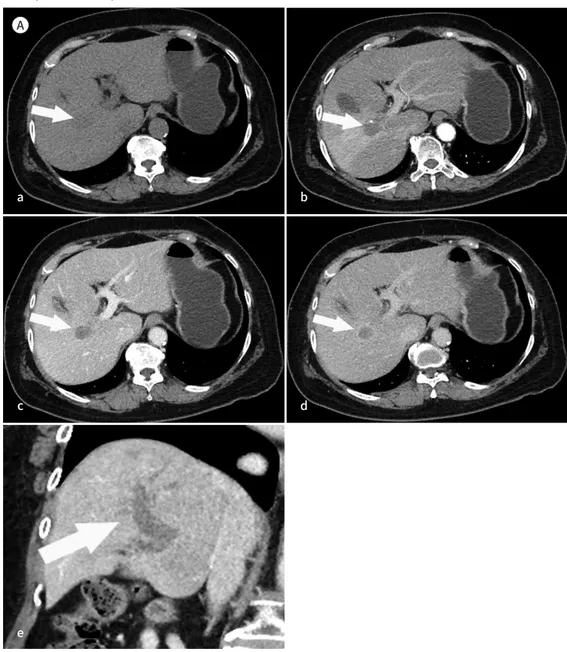
Fig. 1. A 67-year-old woman with lymphoma in the right portal vein.
A. Contrast-enhanced CT images obtained during the pre-contrast phase (a), arterial phase (b), PVP (c), and delayed phase (d). CT images show a hypoattenuated lesion in the right portal vein (arrow, a), with very sub-tle homogenous enhancement (arrow, b and c) in the arterial and PVP. The lesion is enhanced to a lesser de-gree than the liver parenchyma (arrow, b and c) in the arterial and PVP. In the delayed phase, the lesion shows subtle peripheral enhancement (arrow, d). In the coronal image (e), the lesion is clearly seen as a bulging hypoattenuated mass confined to the portal vein (main, right, and left branches, arrow).
PVP = portal venous phases A a c e b d
In our case the portal vein thrombus shows subtle enhancement in the arterial and portal phases and a bulging appearance in CT images and high uptake in 18F-FDG PET CT,
suggest-ing malignancy.
The hepatocellular carcinomas (HCCs) are the most common primary liver malignant tu-mors and frequently cause malignant PVT (2). Other primary tutu-mors for the origin of PVT could be neuroendocrine carcinoma of the pancreas or colon cancer (2). Although rare, there is a case report describing HCC as manifesting only as PVT without a parenchymal mass (5).
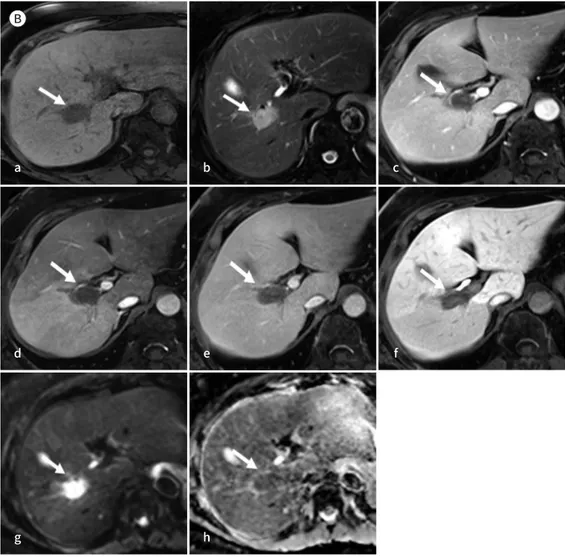
Fig. 1. A 67-year-old woman with lymphoma in the right portal vein.
B. MRI findings of the lymphoma in the right portal vein. The pre-contrast T1-weighted liver image shows a low signal intensity (arrow) compared to that of the liver parenchyma (a), and the T2-weighted image shows a relatively high signal intensity (arrow) (b). Dynamic MRI images after gadolinium-EOB-DTPA en-hancement show very subtle contrast enen-hancement of the tumor thrombus (arrow, c, d, e, and f) during the hepatic arterial (c), portal venous (d), transitional (e), and hepatobiliary (f) (15 minutes delayed) phases rela-tive to the liver parenchyma. Furthermore, the right lobe of the liver shows perfusion disorder caused by portal vein thrombosis in dynamic enhanced images. The right lobe of the liver has a slightly hyperintense signal in the arterial phase, hyperintense signal with clear boundaries in the portal phase, isointense signal in the transitional phase, and hypointense signal sparing the subcapsular area in the hepatobiliary phase. A diffusion-weighted image with b = 800 sec/mm2(g) and apparent diffusion coefficient map (h) show avid
dif-fusion restriction of the lesion (arrow, g and h).
EOB-DTPA = ethoxybenzyl diethylenetriamine pentaacetic acid B a d g h e f b c
In lymphoma, thrombosis occurs quite often, especially in non-Hodgkin’s lymphoma (NHL; the global incidence rate of thrombosis in lymphoma is 6.4% and 6.5% in NHL) (6). However, portal vein tumor thrombosis of lymphoma is extremely rare except in immunocompro-mised patients (6). Regarding the tumor thrombus, only four cases of patients with portal vein lymphoma have been reported and all cases were formed as direct extensions of a domi-nant nodal or extra-nodal mass (1). In some cases, when intravascular large B-cell lymphoma (IVLBCL) concomitantly involves larger vessel such as the portal vein, splenic vein and mesen-teric veins it might be difficult to differentiate with our case radiologically. However in this case report, we exclude IVLBCL since our case does not qualify the definition of IVLBCL (7). IVLBCL is a rare and aggressive form of NHL, and is characterized by the almost exclusive proliferation of lymphoma cells within the lumen of small blood vessels (7). Since our case does not involve small blood vessels but only the portal vein (which is considered a large blood vessel), it is hard to be classified as IVLBCL.
In CT scans, hepatic lymphoma generally shows lower density than the normal hepatic pa-renchyma before contrast enhancement, and lower homogeneous contrast enhancement than the normal hepatic parenchyma after contrast enhancement (8). In MRI, most hepatic lym-phomas show low signal or equivalent signal intensity on T1-weighted imaging, and moderate to high signal intensity on T2-weighted imaging (8). After contrast enhancement, lymphomas
Fig. 1. A 67-year-old woman with lymphoma in the right portal vein.
C.18F-FDG PET CT image (a) and 18F-FDG PET image show high uptake in the right portal vein (b).
D. The cut surface of the resected specimen. The lymphoma is confined to the right portal vein without any other parenchymal lesion.
18F-FDG = fluorine-18 fluorodeoxyglucose
C
a b
typically show low signal intensity in the arterial phase and homogeneous delayed enhance-ment in the PVP, with equivalent signal intensity in the transitional phase (8).
The differential diagnosis between HCC and primary hepatic lymphoma (PHL) is impor-tant. Both HCC and PHL may occur in patients with risk factors, such as cirrhosis and viral hepatitis, show mild signal hyper-intensity on T2-weighted images, and may demonstrate re-stricted diffusion (8). In general, radiologic findings of lymphadenopathy below the level of the renal veins, poor lesion enhancement in all contrast-enhanced phases, and vascular casement without thrombosis favor a diagnosis of lymphoma (8), while arterial phase en-hancement, delayed contrast material washed out with capsular enen-hancement, and vascular thrombosis is more common in HCC (8).
Another differential point is that PVT in HCC patients usually shows a heterogenous appear-ance and disruption of the portal vein walls (9). In our case, however, the PVT shows mild, ho-mogeneous enhancement compared to liver parenchyma without disruption of the portal vein wall. The enhancement pattern with preservation of the vascular structure could be a clue for lymphoma, even though it is extremely rare (8).
Hepatic lymphomas are avidly hypermetabolic in 18F-FDG PET due to hypercellularity, while
most HCCs are not (8). Lesions > 1.5 cm and with FDG accumulation higher than hepatic and splenic FDG are considered positive for lymphoma (10).
Isolated tumor thrombus located only in the portal vein is a rare condition and arriving at a differential diagnosis through imaging can be difficult. However, the significance of treat-ment difference between lymphoma and other primary tumors underscore the importance of keeping lymphoma in the differential diagnosis of a tumor thrombosis in portal vein (1).
Author Contributions
Conceptualization, all authors; data curation, all authors; formal analysis, all authors; investigation, all authors; methodology, all authors; project administration, P.M.; resources, P.M.; supervision, P.M.; visualization, all authors; writing—original draft, all authors; and writing—review & editing, all authors.
Conflicts of Interest
The authors have no potential conflicts of interest to disclose.
REFERENCES
1. Chauhan A, Garg N, Menias CO, Devine CE, Bhosale PR, Balachandran A. Tumor thrombus as a rare presen-tation of lymphoma: a case series of 14 patients. AJR Am J Roentgenol 2015;204:W398-W404
2. Quencer KB, Friedman T, Sheth R, Oklu R. Tumor thrombus: incidence, imaging, prognosis and treatment. Cardiovasc Diagn Ther 2017;7:S165-S177
3. Canellas R, Mehrkhani F, Patino M, Kambadakone A, Sahani D. Characterization of portal vein thrombosis (neoplastic versus bland) on CT images using software-based texture analysis and thrombus density (Houn-sfield units). AJR Am J Roentgenol 2016;207:W81-W87
4. Kim JH, Lee JM, Yoon JH, Lee DH, Lee KB, Han JK, et al. Portal vein thrombosis in patients with hepatocellu-lar carcinoma: diagnostic accuracy of gadoxetic acid-enhanced MR imaging. Radiology 2016;279:773-783
5. Saito M, Seo Y, Yano Y, Uehara K, Hara S, Momose K, et al. Portal venous tumor growth-type of hepatocellu-lar carcinoma without liver parenchyma tumor nodules: a case report. Ann Hepatol 2013;12:969-973
6. Caruso V, Di Castelnuovo A, Meschengieser S, Lazzari MA, De Gaetano G, Storti S, et al. Thrombotic compli-cations in adult patients with lymphoma: a meta-analysis of 29 independent cohorts including 18 018 pa-tients and 1149 events. Blood 2010;115:5322-5328
hepatic portal vein, splenic vein and mesenteric vein tumour embolism. Histopathology 2010;57:648-650
8. Tomasian A, Sandrasegaran K, Elsayes KM, Shanbhogue A, Shaaban A, Menias CO. Hematologic malignan-cies of the liver: spectrum of disease. Radiographics 2015;35:71-86
9. Jha RC, Khera SS, Kalaria AD. Portal vein thrombosis: imaging the spectrum of disease with an emphasis on MRI features. AJR Am J Roentgenol 2018;211:14-24
10. Blechacz B, Gores GJ. Positron emission tomography scan for a hepatic mass. Hepatology 2010;52:2186-2191