Copyright © 2016 The Korean Society of Environmental Health and Toxicology
Introduction
Melanin is a group of natural pigment striking dark color found in organisms like human and animal to protect their skins from ultraviolet (UV) radiation of sun. Melanin is generated by the oxidation of the amino acid tyrosine and considered tissues as melanocytes. Generally, melanin is classified into three types eumelanin, pheomelanin, and neuromelanin. The representa-tive melanin is common eumelanin which shown brown and black color. However, if it is too much UV radiation exposure to skin, it will be made to cover with freckles and even may be lead to skin cancer [1]. In addition, nowadays, there is a trend that people want bright skin from dark skin.
Until now, in order to inhibit to generate melanin in skin, most of the research has been studied through interfering with mela-nin synthesis by inhibiting the tyrosinase [2-4]. In other words, previous studies were designed to methods for treating the mel-anin that has already been generated. Otherwise, some research-ers tried to remove, degradation or treatment directly melanin using by extraction [5,6] extracted from plant or enzymes [7,8]
isolated from cell organelles or themselves [9] after generating it. Proteomic analysis is important skill to find and identify un-known proteins.
Lysosome is a membrane-bound cell organelle having 50 to 60 hydrolytic enzymes associated with antimicrobial [10,11], anti-cancerous [12] and genetic diseases [13]. So, some researchers who are believed to participate in several functions have con-ducted a study with respect to treat melanin using by enzymes in lysosome related organelles [14]. The lysosome is able to ex-tract henʹs egg white [7] and all of eukaryotes like Saccharomy-ces cerevisiae [14] and HeLa cells [8]. In previous studies, it was
conducted research on lysosomal enzymeʹs function.
In this study, we want to know how the lysosome react the melanin. Therefore, it was tried to analysis lysosomal membrane protein isolated from lysosome extracted from activated HeLa cell directly by 2-dimensional electrophoresis (2DE) assay. If the lysosomal membrane proteins are associated with treatment of melanin directly, they will be identified and the revealed pro-teins play important roles to use as a prescription, medical ther-apy and cosmetics to protect UV radiation.
Analysis of lysosomal membrane proteins exposed
to melanin in HeLa cells
Seung Hyuck Bang
1, Dong Jun Park
1, Yang-Hoon Kim
2, Jiho Min
11Department of Bioprocess Engineering, Chonbuk National University, Jeonju; 2Department of Microbiology, Chungbuk
National University, Cheongju, Korea
• Original Article
http://dx.doi.org/10.5620/eht.e2016009eISSN: 2233-6567
Objectives There have been developed to use targeting ability for antimicrobial, antican-cerous, gene therapy and cosmetics through analysis of various membrane proteins isolated from cell organelles.
Methods It was examined about the lysosomal membrane protein extracted from lyso-some isolated from HeLa cell treated by 100 ppm melanin for 24 hours in order to find associated with targeting ability to melanin using by 2-dimensional electrophoresis.
Results The result showed 14 up-regulated (1.5-fold) and 13 down-regulated (2.0-fold) spots in relation to melanin exposure.
Conclusions It has been found that lysosomal membrane proteins are associated with melanin to decolorize and quantity through cellular activation of lysosome.
Keywords Lysosome, Lysosomal membrane proteins, HeLa cell, Melanin, 2-Dimension-al electrophoresis
Correspondence: Jiho Min 567 Baekje-daero, Deokjin-gu, Jeonju 54896, Korea Tel: +82-63-270-2436 Fax: +82-63-270-2306 E-mail: jihomin@jbnu.ac.kr Received: March 23, 2016 Accepted: April 25, 2016 Published: May 4, 2016
Materials and Methods
Cell Cultures and Treatment
HeLa cells were cultured in Dulbecco’s modified Eagle’s medi-um which added 5% newborn calf sermedi-um, 1% penicillin strepto-mycin at 37oC with medium under 5% carbon dioxide. All of
mixtures were exchanged for every two days, and was washed using Dulbecco’s phosphate-buffered saline (DPBS; Gibco, Grnad Island, NY, USA). A 100 ppm melanin was treated when the cells were grown about 70%.
The melanin reagent was purchased from Sigma -Aldrich (St. Louis, MO, USA) and it was dissolved in 0.1 M sodium phos-phate buffer (pH 7.0) to make melanin solution. After that HeLa cells were grown to 70%, they were exposed 100 ppm of melanin solution.
Cell Toxicity Test of HeLa Cells
Harvested cells were separated equivalent amount of them by calculation. When they grow up on the dishes, 100 ppm of mel-anin treated on 0, 6, 12, 24, and 48 hours. Each dish was tested toxicity by 3-(4,5-dimethylthiazol-2-yl)-2,5-diphenyltetrazoli-um bromide (MTT) assay [8]. The colorimetric MTT meta-bolic activity assay is applied to determine whether melanin is able to kill cells. HeLa cells (104 cells/well) were grown on a
96-well plate at 37oC. During exposing melanin on different time,
the supernatant was discarded before washing cells by DPBS. After that, 20 μL of MTT solution (5 mg/mL in DPBS) and 100 μL of medium were supplemented. The plate was then in-cubated for four hours. Finally, 100 μL dimethyl sulfoxide (Sig-ma-Aldrich) was added and the fluorescence signal was mea-sured through enzyme-linked immunosorbent assay reader (Thermo Elctron, Waltham, MA, USA).
Isolation of Whole Lysosomal Membrane Proteins
To extract whole lysosomal membrane proteins in lysosomes, it was isolated lysosomes isolated lysosomes from HeLa cells beforehand. They were rinsed twice with phosphate-buffered saline, and treated with a lysis buffer (20 mM Tris-HCl pH=7.4, 1 mM EDTA, pH 8.8, 1 mM EGTA [Sigma-Aldrich] pH=8.5, 1% v/v Triton X-100 [Daejung Chemicals & Metals Co., Siheung, Korea]), protease inhibitor cocktail (PIC, Sigma-Aldrich), and phenyl methane sulfonyl fluoride (PMSF, Roche, Indianapolis, IN, USA) with ratio 100:1:1 and then vigorous agitation on a vortex mixer and kept in ice for 10 minutes. After incubation on ice, the lysates were centrifuged at 500 rpm, 4oC
for five minutes. The supernatant moved to another tube and centrifuged at 20000×g, 4oC for 30 minutes. Next, lysis buffer
mixed with white pellet including PIC and PMSF. Followed
same vortex mixer and cooling in ice for 10 minutes. And that mixture incubated in ice for 30 minutes to react with lysosome and lysis buffer. After that the lysates were centrifuged at 13000 rpm, 4oC, 10 minutes. The lysosomal membrane proteins were
contained in the pellet. The protein concentration was deter-mined using a Bradford assay with bovine serum albumin as the standard. The pellets were stored at -70oC until analyzed by
2-dimensional gel electrophoresis.
Two-dimensional Polyacrylamide Gel Electrophoresis
Before beginning 2DE assay, whole lysosomal membrane pro-teins isolated from lysosome (20 μg for each sample) in HeLa cells were resoluble into a rehydration buffer containing 350 μL solution (7 M urea, 2 M thiourea, 0.5% v/v Triton X-100, 1% bromophenol blue [Sigma-Aldrich], 35 μL 1 M dithiothreitol [Duchefa Biochemie, Haarlem, Netherlands], and 1.75 μL IPG buffer [GE Healthcare Bio-Sciences, Uppsala, Sweden]) and then the mixture put onto immobiline DryStrip 18 cm, pH 4-7, linear type (GE Healthcare Bio-Sciences, Uppasala, Sweden). A 2.5 mL mineral oil (Bio-Rad, Hercules, CA, USA) was added to cover onto the strip. The rehydration conditions were main-tained at 50 mV for 12 hours at 20oC using a protein isoelectric
focusing (IEF cell, Bio-Rad). After rehydration step, paper wicks were inserted between the IPG strip and each strip holder elec-trode just before isoelectric focusing to adsorb excess water. The isoelectric focusing step was carried out at 20oC at 500 V (2
hours), 1000 V (30 minutes), 2000 V (30 minutes), 4000 (30 minutes), 8000 (until 70000 V), and 500 V (15 minutes). The strips were stored at -70oC until running sodium dodecyl sulfate
poly acrylamide gel electrophoresis (SDS-PAGE). Before per-forming SDS-PAGE, each isoelectric focusing strip was equili-brated for 15 minutes by until 4 mL a solution (2% SDS, 50 mM Tris-HCl pH 8.8, 6 M urea, 30% [v/v] glycerol [Samchun, Py-eongtaek, Korea] 0.5% bromophenol blue, and 100 mg iodo-acetamide [Sigma-Aldrich]). In sodium dodecyl sulfate poly acrylamide gel electrophoresis, the isoelectric focusing strip was loaded onto the 12.5% SDS-PAGE gel (16.8 mL 30% bis/acryl-amide (Bio-Rad), 10 mL of 1.5 M Tris-HCl pH 8.8, 13 mL dis-tilled water (DW), 800 μL of 10 % SDS (Sigma-Aldrich), 800 μL of 10% ammonium persulfate (Sigma-Aldrich), 80 μL tetra-methylethylenediamine (Sigma-Aldrich). Then, approximately 3 mL sealing gel solution (0.5% agarose, 0.2% bromophenol blue) was added onto the surface of the isoelectric focusing strip. The SDS-PAGE separation was performed at 200 mV, 400 mA for six hours at room temperature. Then, IEF gels were fixed with fixing buffer (50% methanol, 12% acetic acid, 38% DW, and 0.00053% formaldehyde) for at least 2 hours.
Silver Staining
After fixing, isoelectric focusing gels were washed three times with 50 % ethanol for 20 minutes. Then, gels were shaking in the sensitizing solution (0.2 g/L, Na2S2O3) for 75 seconds.
Nest, the gels were washed three times with DW by shaking at 100 rpm for 20 seconds prior to react with 2 g/L AgNO3
(Jun-sei Chemical, Tokyo, Japan) solution during 30 minutes. After that, the stained gels were washed two times with DW before developed in a developing solution (60 g/L Na3CO3, 20 mL
0.20 g/L Na2S2O3, 0.00053% formaldehyde [Sigma-Aldrich]).
Usually, it took about five to nine minutes to display all spots on the gel. The developing process was stopped by transferring the gels into the stopping solution (50% methanol, 12% acetic, 38% DW) and shaking at 110 rpm for at least two hours.
Spot Analysis and Data Analysis
All stained gels were scanned to TIF files with a resolution of 300 dpi. The gel images then were cropped to the gel size using Adobe Photoshop CS5 (12.0 version×64 bit, Adobe System Inc., San Jose, CA. USA). Triplicate gels of those used to
visual-ize proteins by silver staining were scanned. The images were evaluated by using Progenesis PG200 version 2006 (Nonlinear Technology, Newcastle upon Tyne, UK) such that all major spots and all changing spots were evaluated the volume of a spot with total volume of all spots in the gels.
Results
Proteomic Analysis of Whole Lysosomal Membrane
Proteins Extracted from Hela Cells Treated by Melanin
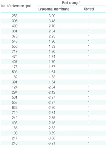
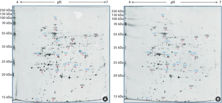
The method of to identify proteins qualitatively, there was used for proteomic analysis using by 2DE assay. It was examined about whole lysosomal membrane proteins extracted from HeLa cells reacted to treatment melanin by 2DE assay. From the proteomic analysis, the reference spots were numbered by Pro-genesis software, and fold change means that expression spots were compared with the control value. Lysosomal membrane proteins extracted from lysosomes in HeLa cells not treated with anything were compared with lysosomal membrane pro-teins treated by melanin. Lysosomal membrane propro-teins ex-tracted from in HeLa cells for proteomic analysis were separated by a pH gradient ranging from 4 to 7 and molecular weight. The separated lysosomal membrane protein spots were profiled after silver staining to distinguish the increased or decreased proteins with control. All of data indicated lysosomal membrane proteins extracted from HeLa cells treated by commercially purchased melanin with 100 ppm for 24 hours. Normal lysosomal expres-sion of lysosomal membrane proteins in Hela cells and were an-alyzed using by Progenesis PG200 software. Based on 2DE gel results, we found a lot of increased and decreased spots com-pared with control gel. Spots were filtered out that the levels of expressed proteins were different for each spot by at least in-creasing 1.5 and dein-creasing 2.0 fold, respectively (Table 1). The 14 spots of up-regulated spots (blue color) over 1.5 fold change and 13 spots of down-regulated spots (red color) down 2.0 fold than control spot change were distinguished (Figure 1). Herein, the up-regulated spots mean that the lysosome membrane pro-teins react more actively to access to the melanin on the con-trary, down-regulated spots do not did. We observed on the ba-sis of results that the differentially expressed proteins were acti-vated by melanin in lysosomes and those lysosomal membrane proteins are possibly associated with quantity of used melanin. For that reason, we tried to investigate the lysosomal membrane proteins reacted to melanin by 2DE assay and found up-regulat-ed 14 spots and 13 down-regulatup-regulat-ed spot in Figure 1. To identify unknown proteins, a lot of researchers used a method which has come close to reveal them through 2DE assay. To get better res-olution spots by proteomic analysis, we used adequate total
Table 1. Lists of the different expressed whole lysosomal membrane pro-teins in HeLa cells in response to melanin with 100 ppm
No. of reference spot Fold change 1 Lysosomal membrane Control
253 3.90 1 396 3.48 1 490 2.70 1 381 2.34 1 370 2.23 1 548 1.90 1 556 1.83 1 717 1.80 1 474 1.74 1 407 1.70 1 175 1.67 1 503 1.64 1 82 1.53 1 391 1.54 1 124 -2.04 1 294 -2.12 1 501 -2.27 1 553 -2.27 1 632 -2.30 1 710 -2.34 1 242 -2.35 1 405 -2.45 1 185 -2.53 1 190 -3.59 1 217 -3.88 1 240 -6.21 1
1Fold change means that expression spots were com pared with the control
tein concentration and then we could gain high level resolution them separated. Furthermore, we can use novel biomarkers found by this research if we identify revealed spots through a matrix assisted laser desorption/ionization time-of-flight mass spectrometry.
Discussion
This study provides the proteomic analysis of lysosomal mem-brane proteins exposed to melanin in HeLa cells. To treat or de-colorize melanin, lysosome has to draw near it. Accordingly, we would like to know how the lysosome approaches the melanin. Because the lysosomes caused to reduce melanin by lysosomal enzymes but it is not possible to contact it without lysosomal membrane having targeting function. In this experiment, we an-alyzed the up-regulated and down-regulated lysosomal mem-brane proteins after exposing 100 ppm melanin by 2 DE. This result showed 14 up-regulated and 13 down-regulated spots. Herein, we classified up-regulated (1.5 fold) and down-regulat-ed (2.0 fold) spots, because it is the most important thing that found up-regulated spots however down-regulated spots will be also regarded to have possibility of sufficiently used. Therefore, our results indicate that lysosomal membrane proteins possibly affect to treat or decolorize the amount of melanin exposed in lysosome due to targeting ability of them. This suggests that if the up-regulated spots use, it is possible to be as biomarkers. And then, it might be helpful in medical fields such as skin can-cer and cosmetics like to decolorize skin.
Acknowledgements
This work was carried out with the support of “Cooperative Research Program for Agriculture Science & Technology De-velopment (project title: DeDe-velopment of Target-specific Anti-microbial and Neutralizing Agents for Livestock Biological Haz-ardous Factors, no. PJ01052701)” Rural Development Admin-istration, Republic of Korea.
Conflict of Interest
The authors have no conflicts of interest associated with mate-rial presented in this paper
ORCID
Seung Hyuck Bang http://orcid.org/0000-0002-9023-6812
Dong Jun Park http://orcid.org/0000-0002-4209-0302
Yang-Hoon Kim http://orcid.org/0000-0002-3406-4868
Jiho Min http://orcid.org/0000-0001-6025-7746
References
1. Kricker A, Armstrong BK, English DR. Sun exposure and non-me-lanocytic skin cancer. Cancer Causes Control 1994;5(4):367-392. 2. Halaban R, Pomerantz SH, Marshall S, Lambert DT, Lerner AB.
Regulation of tyrosinase in human melanocytes grown in culture. J Cell Biol 1983;97(2):480-488.
Figure 1. Whole lysosomal membrane proteins isolated from lysosome in HeLa cells treated by 100 ppm of melanin. (A) Normal lysosomal membrane pro-teins, (B) lysosomal membrane proteins treated by 100 ppm of melanin. Blue color means up-regulated spot and red color means down-regulated spot.
pH pH 4 250 kDa 250 kDa 130 kDa 130 kDa 100 kDa 100 kDa 70 kDa 70 kDa 55 kDa 55 kDa 35 kDa 35 kDa 25 kDa 25 kDa 20 kDa 20 kDa 15 kDa 15 kDa 4 7 7 717 710 717 710 632 632 548 548 503 503 474 474 490 490 501 501 556 556 553 553 396 381407 391 370392 396 381407 391 370392 405 405 190 190 185 185 240 240 294 294 242 242 253 253 217 217 124 124 175 175 82 82 A B
3. Borges CR, Roberts JC, Wilkins DG, Rollins DE. Relationship of melanin degradation products to actual melanin content: applica-tion to human hair. Anal Biochem 2001;290(1):116-125. 4. Butler MJ, Gardiner RB, Day AW. Degradation of melanin or
inhibi-tion of its synthesis: are these a significant approach as a biological control of phytopathogenic fungi? Biol Control 2005;32(2):326-336. 5. No JK, Soung DY, Kim YJ, Shim KH, Jun YS, Rhee SH, et al. Inhibi-tion of tyrosinase by green tea components. Life Sci 1999;65(21): PL241-PL246.
6. Chan EW, Lim YY, Wong LF, Lianto FS, Wong SK, Lim KK, et al. Antioxidant and tyrosinase inhibition properties of leaves and rhi-zomes of ginger species. Food Chem 2008;109(3): 477-483. 7. Bang SH, Kim P, Oh SJ, Kim YH, Min J. Impact of solvent pH on
direct immobilization of lysosome-related cell organelle extracts on TiO2 for melanin treatment. J Microbiol Biotechnol 2015;25(5): 718-722.
8. Park DJ, Sekhon SS, Ahn JY, Yoon H, Lee L, Ko JH, et al. Different expression patterns of lysosomal proteins exposed to melanin in HeLa cells. Toxicol Environ Health Sci 2015;7(5):272-276.
9. Otaki N, Seiji M. Degradation of melanosomes by lysosomes. J In-vest Dermatol 1971;57(1):1-5.
10. Bang SH, Jang A, Yoon J, Kim P, Kim JS, Kim YH, et al. Evaluation of whole lysosomal enzymes directly immobilized on titanium (IV) oxide used in the development of antimicrobial agents. En-zyme Microb Technol 2011;49(3):260-265.
11. Yoon J, Park JM, Kim KJ, Kim YH, Min J. Antimicrobial activity of the cell organelles, lysosomes isolated from egg white. J Microbiol Biotechnol 2009;19(11):1364-1368.
12. Linder S, Shoshan MC. Lysosomes and endoplasmic reticulum: targets for improved, selective anticancer therapy. Drug Resist Up-dat 2005;8(4):199-204.
13. Futerman AH, van Meer G. The cell biology of lysosomal storage disorders. Nat Rev Mol Cell Biol 2004;5(7):554-565.
14. Yoon J, Kim YH, Ahn JY, Lee HC, Oh SJ, Chung BW, et al. Melanin reduction by peroxidase activity in lysosome-related organelle ex-tracts from hen egg whites, HeLa cells, and Saccharomyces cerevi-siae. Mol Cell Toxicol 2015;11(4):441-447.